A corneal ulcer is an abrasion or defect in the clear surface tissue of the eye. Ulcers may be shallow or deep. As this is a painful condition, the signs that are usually seen are squinting and tearing. Many ulcers are caused by trauma — possibly through an interaction with another cat or dog, or by something lodged under the eyelid, particularly foxtails. Ulcers can also occur due to “rolling in” of the eyelids (entropion) causing the lashes to rub on the surface of the eye, extra eyelashes (ectopic cilia), or inadequate tear production (dry eye). There are two types of internal degeneration of the cornea that can lead to corneal ulceration.
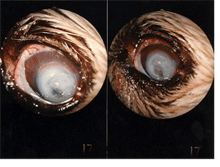
Corneal dystrophy is characterized by abnormal deposits within the cornea which usually contain a combination of calcium and fats (cholesterol) within the superficial and deep layers. These may occur in one or both eyes and often have no apparent cause, although they may be associated with low thyroid levels (hypothyroidism). They appear as white, granular spots which usually do not interfere with vision. When initially seen, the spots are monitored for 1-2 months. If there is evidence of progression during that time, eye drops can be used to bind calcium and thus, slow the progression of the opacity. in rare cases. usually in dogs over 12 years old, affected eyes may become suddenly painful with ulceration occurring as a reaction to the abnormal material in the cornea. Once ulceration occurs surgery is necessary to remove the deposit. If untreated, the ulceration may progress, even resulting in rupture of the eye in the most severe cases.
Defective cell adhesion is another cause of superficial ulcers in middle-aged to older dogs. This condition appears to be caused by abnormal cell “cement” so that the surface cells fall off because they can’t stick to each other. Treatment is is directed at stimulating the cells to produce new cement. Initially, the cornea is debrided to remove the unattached cells. A collagen shield may be applied to the eye. These shields look very much like soft contact lenses; once applied, they adhere to the eye and dissolve over approximately four days. A large plastic collar is used to prevent rubbing at the eye which could dislodge the shield. In one week most cases have resolved; those that persist usually require surgery (superficial keratectomy) using an operating microscope to remove the unhealthy tissue under general anesthesia. This procedure results in resolution in nearly all cases. Since the condition is caused by abnormal tissue structure, recurrence is possible (although rare) in the same or the other eye.
ATLANTA VETERINARY EYE CLINIC
33 Avondale Plaza North
Avondale Estates, GA 30002
Map & Directions | Email
OFFICE HOURS
M W F 9:00 am - 3:00 pm
T Th 9:00 am - 6:00 pm
Saturdays Closed
Sun Closed
OFFICE HOURS
CALL TO SCHEDULE AN APPOINTMENT
WE ACCEPT